Suphaket Saenthaweesuk1,
Narongsuk Munkong2,
Wason Parklak1,
Atcharaporn Thaeomor3,
Janeyuth Chaisakul4,
Nuntiya Somparn1 ![]()
For correspondence:- Nuntiya Somparn Email: nuntiya_tom@hotmail.com Tel:+6629269710
Received: 4 July 2016 Accepted: 5 December 2016 Published: 31 January 2017
Citation: Saenthaweesuk S, Munkong N, Parklak W, Thaeomor A, Chaisakul J, Somparn N. Hepatoprotective and antioxidant effects of Cymbopogon citratus Stapf (Lemon grass) extract in paracetamol-induced hepatotoxicity in rats. Trop J Pharm Res 2017; 16(1):101-107 doi: 10.4314/tjpr.v16i1.13
© 2017 The authors.
This is an Open Access article that uses a funding model which does not charge readers or their institutions for access and distributed under the terms of the Creative Commons Attribution License (http://creativecommons.org/licenses/by/4.0) and the Budapest Open Access Initiative (http://www.budapestopenaccessinitiative.org/read), which permit unrestricted use, distribution, and reproduction in any medium, provided the original work is properly credited..
Purpose: To investigate the protective effect of Cymbopogon citrates Stapf. (CS, lemongrass) extract on paracetamol (PCM)-induced hepatotoxicity in rats.
Methods: The rats were orally administered CS extract (1000 mg/kg/day) for 30 days prior to induction of hepatotoxicity by a single oral administration of PCM (3 g/kg). Hepatoprotection was assessed by measuring the level of hepatic markers including aspartate transaminase (AST), alanine transaminase (ALT) and oxidant/antioxidant markers including Malondialdehyde (MDA), protein carbonyl, and glutathione (GSH) in liver homogenate and serum. Phytochemical screening of the CS extract was also performed.
Results: Phytochemical screening of the extract indicate the presence of tannins, flavonoids, and phenolic compounds. Elevation of serum AST, ALT, and MDA levels along with depletion GSH in the liver were observed in rats treated with PCM alone compared with control (p < 0.05). Pre-treatment of the animal with CS extract reduced the levels of hepatic markers (AST and ALT). Pre-treatment with CS extract also significantly reduced oxidative stress induced by PCM as shown by an increase in GSH level and reduction of MDA compared to rats treated with PCM alone (p<0.05).
Conclusion: The results indicate that CS possesses antioxidant activity and it exerts its effect by reducing lipid peroxidation and restoring GSH. Pre-treatment with CS extract reduces oxidative stress and ameliorates hepatic injury induced by PCM.
Introduction
Paracetamol (PCM) is an over-the-counter drug commonly used for its analgesic and antipyretic activities. At therapeutic doses, PCM is considered safe. However, PCM overdose can cause severe hepatotoxicity and nephrotoxicity. PCM is activated and converted by liver enzymes to N-acetyl-p-benzoquinoneimine (NAPQI), a toxic metabolite that can be detoxified by glutathione (GSH), which results in extensive hepatic GSH depletion and oxidative stress [1]. GSH depletion leads to lipid peroxidation, which further leads to the initiation of liver damage. Several studies have demonstrated that the antioxidant properties of certain medicinal plants, such as ginseng, soybean, and pomegranates, can help provide substantial protective and health benefits against oxidative stress [2,3].
Cymbopogon citrates Stapf. (CS), commonly known as lemongrass, is a widely used herb in Southeast Asia. Studies have reported the pharmacological properties, including free radical scavengers, anti-oxidant, anti-inflammatory and anti-mutagenicity properties, of specific parts of CS [4]. Moreover, the hypoglycemic and hypolipidemic effects of fresh CS leaf extracts have been demonstrated [5]. Therefore, the present study investigates the antioxidant potential of CS against PCM-induced hepatotoxicity in rats.
Methods
Chemicals and drugs
Paracetamol, 2, 2-dipheneyl-1-picrylhydrazil (DPPH), thiobarbituric acid (TBA) were purchased from Sigma Chemical Company (St. Louis, MO, USA). All other chemicals and reagents used were of analytical grade.
Plant material and identification
Fresh CS was procured from a local agricultural field in Khon Kaen Province, Thailand in November 2014. The plant was authenticated by Ms. Dujhathai Anekchai, a taxonomist from the Faculty of Pharmaceutical Sciences, Prince of Songkla University, Songkla, Thailand. A voucher specimen (no. SKP 081030301) was deposited in the herbarium of the same institution.
Extraction and phytochemical screening
Whole plants were washed thoroughly in water, cut into small pieces, soaked in 50 % ethanol (1 part of plant material: 2 parts of 50 % ethanol) for 72 h, and then filtered. The filtrate was concentrated using rotary vacuum evaporation and then lyophilized with a freeze dryer. The yield of the extract was 1.86 %. The extract was kept at −80 °C before its screening for the presence of tannins, flavonoids, and phenolic compounds [6].
Total phenolic content assay
The CS extract was assayed for total phenolic content using the Folin-Ciocalteu reagent according to a previously described method [6]. Gallic acid was used as a standard, and the results were expressed as gallic acid equivalent per gram CS extract.
1,1-Diphenyl-2-picrylhydrazyl (DPPH) radical scavenging assay
The DPPH radical-scavenging activity was determined as per the previously described method [7]. Briefly, 0.08 mM DPPH was prepared in 100 % ethanol. DPPH solution, Tris buffer, and 80 % ethanol were mixed in order to obtain a 1:1:1 ratio for 1.8 mL. Then the plant extract (0.6 mL) was added and incubated for 30 min in the dark. The absorbance was read at 525 nm.
Animals and experimental design
The animals were divided into 6 groups, each of which consisted of 6 rats that were treated as follows:
Group I: (Control) distilled water (DI)
Group II: (PCM only) DI + PCM
Group III: (positive control) DI + PCM followed by N-acetylcysteine (NAC).
Group IV: DI + PCM followed by CS extract
Group V : CS extract
Group VI: CS extract followed by PCM
DI (1 mL/kg) and CS (1,000 mg/kg) were administered orally once daily for 30 consecutive days, and then, the animals were administered with a single oral dose of PCM (3 g/kg) on the 31st day. Groups III and IV received NAC (1 g/kg) or CS (1,000 mg/kg) orally after the single oral dose of PCM.
At 24 h after PCM administration, all the animals were anesthetized and euthanized. Blood was collected by cardiac puncture, and the liver tissue was dissected for biological and histological evaluation. The protocol for the present study was approved by the Animal Research Committee of Thammasat University, Thailand (license no. AE 005/2015). All institutional and national guidelines for the care and use of laboratory animals were followed in this study [8].
A single dose of PCM was selected according to the previous reports in the hepatoxicity-induced rat model [9,10]. The dose of extract (1000 mg/kg) used in the present study was selected based on our preliminary study on serum total antioxidant capacity (TAC). Serum TAC, which is considered as the cumulative action of all the antioxidants, was demonstrated to be significantly elevated only in 1000 mg/kg CS treated rat (1.74 ± 0.45 mM trolox equivalent) compared to the control group (1.05 ± 0.75 mM trolox equivalent). However, the doses of 250 mg/kg and 500 mg/kg (1.15 ± 0.77 and 1.10 ± 0.87 mM trolox equivalent, respectively) did not demonstrate the significant changes in the serum TAC.
Acute toxicity studies
Thirty-six male Sprague-Dawley rats, weighing 200 - 250 g, were used for the acute toxicity study. They were allowed to acclimatize for seven days to the laboratory condition before the experiment. The procedure was performed according to OECD 425 guidelines [11], with respect to the guidelines that the volume of administration should not excess 1 mL/100 g body weight. The maximum concentration of CS dissolved in aqueous solution is 1000 mg/mL. Therefore, our limit test dose is 2000 mg. The rats received 2,000 mg/kg CS extract orally. On testing the first dose with one animal, we found that the animal survived. Thus, the remaining animals (n = 4) were administered with 2,000 mg/kg CS extract. They were continuously observed for 48 h to detect the changes in autonomic or behavioral responses. Any mortality or behavioral changes during the experimentation period of 15 days were also recorded. All the animals survived during the study period, and we could not increase the test dose due to the limits of the dissolution. We, thus, anesthetized and euthanized the animals. Blood was collected by cardiac puncture, and liver tissue was dissected out for biological and histological evaluation. The LD50 > 2000 mg/kg body weight may be assumed.
Evaluation of antioxidant activity
Malondialdehyde (MDA), a marker of lipid peroxidation, was assayed in plasma as thiobarbituric acid reactive products (TBAR) [12]. In brief, 150 ml of plasma was reacted with 125 mL of 10 % TCA, 125 mL of 5 mM EDTA, 125 mL of 8 % SDS, and 10 mL of 0.5 mg/mL of an antioxidant, butylhydroxytoluene (BHT), to prevent auto-oxidation during the assay. The mixture was left for 10 min, then 435 mL of 0.6 % thiobarbituric acid (TBA) was added, and the mixture was heated for 30 min in a boiling water bath. After cooling to room temperature, 10,000 g of the mixture was centrifuged at 25 °C for 10 min. The absorbance of the supernatant was measured at 532 nm. A standard curve of MDA was generated by using appropriate concentrations of standard 1,1,3,3-tetraethoxypropane (TEP) (0.3 - 10 mmol/L).
Serum protein carbonyl was assessed with protein carbonyl content assay kit (MAK094, Sigma Chemical Company (St. Louis, MO, USA)) while the GSH content in liver was assessed with glutathione assay kit (CS0260, Sigma Chemical Company (St Louis, MO, USA).
Measurement of liver and kidney function markers
The blood of animals from both acute toxicity test and hepatoprotective study was collected by cardiac puncture. 10 mL of whole blood was allowed to clot in a conical tube at room temperature. After coagulation, the blood was centrifuged at 2,000 rpm 4 °C for 10 min to separate the serum. The levels of liver function markers, including kinetic assay of enzyme aspartate aminotransferase (AST) and alanine aminotransferase (ALT), and kidney function markers, including blood urea nitrogen (BUN) and serum creatinine (Cr), were measured using automated machine analyzer at The Clinical Pathology Laboratory Unit, Thammasat University Hospital.
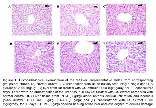
Histopathological examination of rat liver
All animals from the acute toxicity test and the hepatoprotective study were anesthetized and euthanized. The liver tissue was dissected out, washed in ice cool PBS pH 7.4, fixed in 4 % paraformaldehyde, and embedded in paraffin. The serial sections were cut 5 mm thick and stained with hematoxylin-eosin (H&E) and examined under a photomicroscope.
Statistical analysis
Data are presented as mean ± SEM. Analysis of variance (ANOVA) with Duncan post-hoc test was used to determine significant differences between each experimental group. An ANOVA on rank test was also performed for the non-parametric test. The level of significance was set at p < 0.05 using SigmaStat software version 4 (Systat Software, Inc. California, USA).
Results
Antioxidant properties
The CS extract contain several chemicals with antioxidant properties including polyphenolic compounds. The extract contained phenolic compounds 1,400.10 ± 0.47 mg of gallic acid equivalents per gram CS extract. The free radical scavenging activity assessed by DPPH assay gave IC50 of 168.77 ± 3.32g/mL, which is relatively lower than that of BHT with IC50 of 12.34 ± 1.14 µg/mL.
Acute toxicity of the CS extract
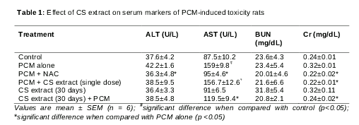
After receiving a single dose of 2,000 mg/kg, the rats showed no mortality or any clinical signs during the experimental period. In addition, treatment with a single dose of CS extract did not affect the serum markers of liver and kidney function. The serum AST, ALT, BUN, and Cr levels were normal (), and no abnormality of the liver tissue was observed (). It may be assumed that the LD50 of CS is > 2000 mg/kg.
Effect of CS extract on the serum markers of liver and kidney function
Administration of the CS extract alone for 30 days did not affect liver and kidney function as shown by the normal serum AST, ALT, BUN and Cr levels in rats group V (). No abnormality of the liver tissue was observed in any rat treated with CS extract for 30 days. These findings indicated that the CS water extract administration in rats was generally safe even with at a dose up to 1,000 mg/kg/day for 30 consecutive days.
Treatment of rats with PCM alone showed a significant increase in serum ALT and AST levels compared with normal control rats, indicating that the hepatotoxicity was induced by PCM. Pre-treatment with 1,000 mg/kg/day of the CS extract for 30 days or treatment with NAC significantly reduced the elevated serum AST and ALT levels (p < 0.05) (Table1). The results indicated a hepatoprotection of CS extract against the hepatotoxic effect of PCM.
Effect of CS extract on oxidant and antioxidant status
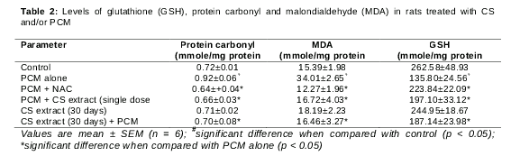
The serum MDA level was measured as a marker of lipid peroxidation, and protein carbonyl was measured as a marker of protein oxidation. It was found that MDA and protein carbonyl levels were significantly elevated in animals treated with PCM compared to control animals (p < 0.05) (). Lipid peroxidation and protein oxidation levels were reduced by pre-treatment with the CS extract. In addition, depletion of liver GSH was found in rats treated with PCM compared with control (p < 0.05). Pre-treatment with CS extract showed an elevation of liver GSH content compared to the PCM treated alone ().
Effect of CS extract on liver histology
When compared with control rats, the liver section of PCM-treated rats exhibited cellular infiltration and necrosis. Pre-treatment with CS extract showed healing of the liver and less degree of cellular damage, whereas the treatment with NAC can prevent the cellular damage ().
Discussion
PCM is the most common drug used for pain and fever. Generally, it is safe at the recommended dose; however, its long-term usage or abuse by taking high dose can induce hepatocellular damages or necrosis [13]. Hepatotoxicity of PCM has been attributed to the formation of toxic metabolites called NAPQI, which is activated by hepatic cytochrome p450. Initially, NAPQI is detoxified with reduced glutathione (GSH).
However, when the rate of NAPQI formation exceeds the rate of detoxification by GSH, it oxidizes tissue macromolecules such as lipid or sulfhydryl group of proteins. GSH depletion subsequently leads to increased lipid peroxidation by abstracting hydrogen from polyunsaturated fatty acid, which ultimately damages liver tissue [13,14].
One of the end-products of lipid peroxidation is MDA. The increase in MDA level in liver, which can be induced by PCM intake, suggests enhanced lipid peroxidation and is an indicator of hepatic tissue damage [14,15]. Beside the depletion of glutathione, NAPQI can exert initial cell stress through a wide range of mechanisms including binding to enzymes, lipids, nucleic acids, and other cell structures [16]. After cellular damages, hepatic enzymes including AST and ALT are released into circulation. A significant elevation of AST and ALT levels is, thus, often found in PCM intoxication with hepatocellular injury [17,18].
In the present study, treating rats with high-dose PCM alone resulted in the significant increases in AST, MDa, and protein carbonyl, and a significant decrease in GSH level. It is interesting to discover that rats treated with CS extract (both PCM followed by single dose of CS and 30-day CS followed by PCM) had a significant reduction of lipid peroxidation and resumption of GSH level compared to rats treated with PCM alone. The degree of liver tissue damage as measured by the level of AST enzyme was also significantly improved among rats treated with CS extract compared to rats treated with PCM alone. These results are consistent with the histopathological findings of the liver tissue from the tested rats.
CS is an edible herb commonly used worldwide, and its safe consumption is historically appreciated. Consumption of CS water extract for 30 days in our study showed its safety profile in the experimental animals at the dose up to 1000 mg/kg/day. In line with other studies, we found that lemongrass contains alkaloids, volatile and non-volatile terpenoids, flavonoids, carotenoids, and tannins [19]. We also demonstrated the antioxidant activity of the CS extract. This radical scavenging activity and antioxidant effect could be due to the presence of one or more active principles in the plant.
Several studies showed that plants extracts containing antioxidants exhibit the hepatoprotective activity through the regulatory action on cellular permeability, stability, and suppressing oxidative stress [20-22]. Certain flavonoids have been demonstrated to have a protective effect on liver attributable to their antioxidant properties [20-22]. Tannins, which have potent antioxidants and anti-inflammatory activities, have been reported to reduce the levels of lipid peroxide in liver cells and thus, prevent hepatocytes from the destructive effects [23]. Phenolic compounds were also reported as the important active ingredients of natural products that are responsible for the antioxidant effect [24-27]. Findings from our phytochemical analysis showed that the water extracts of CS contained phenolic compounds and tannins. It is likely that these compounds contribute to the hepatoprotective effects of the extract. However, the complete phytochemical constituents of the CS extract, and their mechanism of action needs further investigated.
Conclusion
The results suggest that treatment with CS water extract exhibits antioxidant effect by reduction of lipid peroxidation and induction of GSH levels, thereby, ameliorating the liver injury induced by PCM.
Declarations
Acknowledgement
References
Archives


News Updates